Hospital

Introducing you : the heart!
Well, and lungs too.
This is an emergency open thoracotomy (surgical opening of the chest cavity, or thorax) that is performed upon the anterior chest wall.
It is performed by surgeons to gain access to the thoracic organs, most commonly the heart, the lungs, or the esophagus, or for access to the thoracic aorta or the anterior spine (the latter necessary in case of tumors in the spine). A resuscitative or emergency thoracotomy may be performed to resuscitate a patient who is near death as a result of a chest injury. It provides access to the chest cavity to control injury-related bleeding from the heart, cardiac compressions to restore a normal heart rhythm, or to relieve pressure on the heart caused by cardiac tamponade (accumulation of fluid in the space between the heart's muscle and outer lining).

Sleeve gastrectomy! This is the portion of the stomach that was removed during a laparoscopic sleeve gastrectomy, a type of bariatric (surgical weight loss) procedure.
About 75% of the stomach was removed by a surgical resection of a large portion of the stomach along the greater curvature, leaving a narrow gastric “tube” or “sleeve”. No intestines are removed or bypassed during the sleeve gastrectomy.
The basic principle behind that procedure is that it greatly reduces the size of your stomach and limits the amount of food that can be eaten at one time. It does not cause decreased absorption of nutrients or bypass your intestines and after eating a small amount of food, you will feel full very quickly and continue to feel full for several hours.

Six-pack tumors!!! This photo shows a fibrosarcoma of the rectus abdominis muscle in a 30 year-old patient. He was asymptomatic with large painless mass palpated lateral to the umbilicus originating from the left rectus abdominis muscle.
Fibrosarcomas are a type of malignant soft tissue neoplasms that is composed of malignant and immature proliferating fibroblasts in a collagen background with a strong tendency to recur locally after resection. They are of mesenchymal cell origin in which histologically the predominant cells are fibroblasts that divide excessively without cellular control.
Operation was done with a total excision of the mass and removal of the rectus abdominis muscle to avoid local recurrence.

This patient was discovered to have gallstone ileus, a rare complication of cholelithiasis which has a high rate of mortality. An unusual multi-step process must occur in order to cause this complication.
Gallstones of this size (typically > 2cm) can enter the bowel following the development of a fistula between the gallbladder and the small bowel. These fistulas can be asymptomatic, and the first signs of illness are usually related to intestinal obstruction.
The optimal surgical procedure shown here is enterotomy with stone extraction, which appears to be associated with better outcomes than more invasive techniques.
Case contributed by Figure1.

This eye is literally watching him...
This is an eye enucleation after death, which is the removal of the eyeball that leaves the eye muscles and remaining orbital contents intact.
This is done for the purpose of donation, the most common is the cornea, which can be harvested up to twelve hours after death, but ideally within six hours. Eye banks are the institutions responsible for collecting (harvesting) and processing donor corneas, and for distributing them to trained corneal graft surgeons.
Cornea harvesting is the surgical removal from a deceased person of either the whole eye (enucleation) or the cornea (in situ corneal excision) for different medical uses in order to improve visual acuity, to reconstruct and to improve the appearance of patients with corneal scars that have given a whitish or opaque hue to the cornea.
Photo credit - @joaquinmac

Actual blood vessels of a person who donated their body for scientific display purposes! Science is fascinating isn't it?
The principle by which preservation of the blood vessels is done is pretty simple, injection of a solution, more precisely - plastic. An incision is made in a vein in the foot, then injection of the plastic into a vein in the neck - the blood gets pushed out the foot, and the plastic fills out the rest of the vessels, which harden them and keeps them safe while dissolving everything else with an acid.

Here's a resected stomach after sleeve gastrectomy of about 80% of the stomach.
This is a surgical weight-loss procedure in which the stomach is reduced to about 20-25% of its original size, by surgical removal of a large portion of the stomach along the greater curvature. The result is a sleeve or tube like structure, a smaller stomach which is about the size of a banana. It limits the amount of food you can eat by making you feel full after eating small amounts of food.
The surgery is a minimally invasive surgery, generally performed laparoscopically.
Photo credit : @rabee3s

Oh snap!
Finger injuries are common and range from minor cuts and scrapes to wounds with major damage to bone, tendons, and ligaments.
This finger injury isn't easy to treat because it shows multiple tissue damage, including the dislocation of the joint that causes a bone to move out of its normal alignment with another bone, ligament injury (sprain), tendon injury and probably nerve injury that causes numbness (loss of sensation). A finger may be injured by a direct blow or cut and many are work-related during which the finger is jammed, twisted, or stretched.

A hematoma organized on the back of hand with skin necrosis after puncture in anticoagulated patient.
This patient suffered from a medication-induced coagulopathy. Patients typically receive these medications for chronic antithrombotic therapy in the prevention of thrombosis that can lead to stroke or myocardial ischemia, thromboprophylaxis following surgery, or treatment of acute thromboembolism or coronary syndrome. The intensity and duration of anticoagulation affect the risk of spontaneous, as well as procedural-related bleeding complications and hematoma formation.
Photo by : @cpmantrana

A close-up of the frontal lobe, so close that we can almost see its thoughts!
Photo credit : @rpaglioli

Those twin fetuses have been spontaneously aborted during 20th weeks gestation due to medical reasons.
Miscarriage is an unfortunate thing to deal with, and many medical complications during pregnancy can lead to it such as an infection, uterine and hormonal abnormalities, unhealthy lifestyle, drug and alcohol usage and smoking, exposure to environmental and workplace hazards, immune system problems, and list goes on and on. In addition, women may be at increased risk for miscarriage as they age.
When a miscarriage occurs, the tissue passed from the vagina should be examined. This is done to determine if it was a normal placenta or a hydatidiform mole (a rare growth that forms inside the womb early in pregnancy). It is also important to determine whether any pregnancy tissue remains in the uterus.
Surgery (suction curettage, D&C) or medicine may be needed to remove the remaining contents from your womb. After treatment, women usually resume their normal menstrual cycle within 4 to 6 weeks.
Photo sent by : @saeedhajizadehh

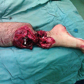
Traumatic leg injury as a result of a motorcycle accident.
Injuries to the legs and feet are fairly easy to diagnose and for the most part, they' re fairly superficial, but some are just the opposite.
Leg injuries due to motorcycle accidents are becoming increasingly common, and a relatively high proportion of these injuries are severe, requiring intensive and prolonged treatment with an often disappointing final result.
Photo credit : @hamiidreza_m1

Demonstrating how big is that colon. Really big.

ight mastectomy to treat breast cancer.
Mastectomy means the surgical removal of the breast, either completely or partially.
This is the standard treatment for breast cancer. There are many subtypes to the procedure such as radical and partial mastectomies, lumpectomy (breast-conserving surgery), and quadrantectomy. The decision regarding to which procedure should be taken is depending on the number of lesions and their aggressiveness, the breast size, and availability of radiation and other therapies.
Newer mastectomy techniques can preserve breast skin and allow for a more natural breast appearance following the procedure. This is known as skin-sparing mastectomy.
Photo by : @nazandedeogluu

An infant with sirenomelia, also known as the mermaid syndrome, undergoing a surgical repair for joined legs.
This is a very rare congenital malformation in which the legs are fused together, giving them the appearance of a mermaid's tail.
The condition is fatal and incompatible with life because of complications associated with abnormal kidney and urinary bladder development and function.
exact cause of sirenomelia is unknown and heavily debated, but it is believed that it results from a failure of normal vascular supply from the lower aorta in utero, causing severe ischemia to the caudal portion of the fetus.
Surgery has been successful in separating joined legs, requiring the coordinated efforts of a team of specialists such as pediatricians, surgeons, cardiologists, orthopedists, and kidney specialists (nephrologists). In preparation for surgery, balloon-like tissue expanders are inserted under the skin. When they are filled with a salt solution over a period of time, the balloons expand making the skin stretch and grow. The excess skin is then used to cover the legs once they are separated.

"Hearts will never be practical until they are made unbreakable". This is what our heart looks like, from the moment it begins beating until the moment it stops, the human heart works tirelessly. It pumps blood and nutrients throughout our body's different systems, and like a pumping machine, the heart provides the power needed for life.

Temporal lobe epilepsy surgery in a 24-year-old female patient!
An intraoperative electrocorticography (ECoG) is used with recording strip electrodes placed on the exposed cortex of the right temporal lobe.
Photo credit :@rpaglioli

Surgeon holds a donor kidney right before transplantation.
The whole operation was live-tweeted and shared by UWMEdicineKidney right from the OR. The donor kidney has been chilling in an ice bath, and the scrub nurse was holding the container as the doctor removed the kidney, holding it out for a photograph.
Kidney transplants are the most common transplant operations, and are the best treatment option for an end-stage renal disease, in which kidney failure is so advanced that it cannot be reversed or treated with medications. Interesting fact is that in some kidney transplants, the old kidney isn't removed. It is essentially disconnected and then pushed into a safe spot in the body, unless they are causing medical problems like high blood pressure, infections, or are too large to stay in the body.

ell that is one super duper huge enormous mega colon!
A colectomy (surgical removal of the entire colon) is usually indicated for the treatment of a complicated toxic megacolon in case other treatment methods aren't benefiting, as seen here.

Let's have a brain dissection and discussion!
Brain lobectomy is usually the surgical treatment for epilepsy. Patients with epilepsy, whose seizures arise commonly from the temporal or frontal lobes, have a high probability that their seizures will not be controlled with anti-seizure medications alone. Surgery for epilepsy is a well-established procedure with excellent results.
Temporal lobectomy is a surgical procedure designed to remove seizure causing brain tissue. The temporal lobes are the brain segments located on either side of the head just above the ear. During surgery, craniotomy is performed and a small, seizure-causing portion of the temporal lobe is removed, using an intracranial EEG monitoring during the surgery to help the surgeon pinpoint the exact location of the areas of the brain causing the seizures.
Photo credit : @nazandedeogluu

Bilateral self-enucleation of eyes! Enucleation simply means the removal of an entire structure (such as an eyeball or tumor), without rupture of its enveloping cover or sac.
Self-enucleation of eyes in an extreme but fortunately rare form of self-harm. This 48-year-old male was brought to the Emergency Department with a history of self-gouging of both his eyes. Both his orbits were bandaged and there was very little ooze down over cheeks. His enucleated eyeballs, along with a long stump of optic nerve, were stored in a pot filled with normal saline (above figure). He was calm and apparently in no pain. The history was patchy and his relatives said that he indeed had a very troubled family life. There was no significant past ocular history. He was a known epileptic and had a recent epileptic attack, prior to the self-enucleation. A psychiatric consult concluded that he was suffering from a postictal psychosis, which had led him to do such an act.
The next morning the sockets were examined closely and it was decided to leave them to heal spontaneously. Chloramphenicol eye ointment was prescribed for the sockets. He was registered fully blind and referred to artificial eye centre for prosthesis.

A healthy donor liver transplanted to a cancer patient who had liver metastases that has spread from another organ affected by the cancer.
Almost any primary cancer can spread to the liver, among them are breast, colon, rectum, esophagus, lung, skin, pancreas, stomach and uterus. In some countries, as many as half of colorectal cancers are spreading to other organs, they metastasise, usually to the liver. A liver transplantation is done because the spread of tumors is often so pervasive that the surgical removal of affected part of the liver is not a viable option. Patients can count on some relief from chemotherapy, but the results are generally insufficient.

Interesting case of an intraosseous hemangioma of the frontal bone!
These are rare bone (osseous) tumors that constitute less than 1% of all bone tumors. They're classified as benign, slow-growing tumors of vascular nature (hence the name hemangioma), that originate and expand inside bone structures.
Although found in any bone, seventy-five percent of intraosseous hemangiomas are located in the vertebral bodies (especially the thoracic spine), skull (involved outer table, normal inner table), and facial bones.
Vertebral body hemangiomas are usually asymptomatic and left untreated. Sometimes they are incidentally found in a patient with low back pain. They may develop a soft tissue mass which may lead to neurologic symptoms like pain and numbness.
A CT or MRI is used to diagnose and define the epidural extent.
Photo credit : @brainpage

A single photo can tell a thousand different stories. This breathtaking photo is certainly one of them.

Severe injury to the arm involving structures deep beneath the skin including muscles, bones, nerves, and blood vessels as a result of a pitbull bite.
Never piss off an angry dog. Pitbulls are widely considered as one of the most dangerous dog breeds. It has a bite force of 235 pounds, which is roughly 75% of the bite force of a Rottweiler. But, despite its negative image, If properly trained and socialized, these dogs can be very loyal friends. In fact, many other dog breeds can cause the same damage, if the owner push them to do so.
Photo credit @cpmantrana

Coronal approach to the upper facial skeleton.
This fascinating image shows the coronal or bitemporal incision which is a surgical approach to the regions of the facial skeleton, including the zygomatic arch. Provides excellent access to these areas with minimal complications, with the major advantage being that in this approach most of the surgical scar is hidden within the hairline.
The variety of cases in which it has proven to be useful include craniofacial reconstruction, facial trauma, and tumor resection.
Photo credit : @maxillofacialtips

This is a rare condition of an acute anterior uveitis in a patient with ankylosing spondylitis.
Ankylosing spondylitis is an inflammatory disease that can cause some of the vertebrae in your spine to fuse together. This fusing makes the spine less flexible and can result in a hunched-forward posture.
So what uveitis has to do with this?
HLA (or Human Leukocyte Antigen) halotype association with different diseases, in this case HLA-B27.
The HLA system is the locus of genes that encode for proteins on the surface of cells that are responsible for regulation of the immune system in humans. People with certain HLA antigens are more likely to develop certain autoimmune diseases, with more than 100 diseases already associated with different alleles of HLA gene.
A classic example is that HLA-B27 allele increases the risk of developing an inflammatory joint disease called ankylosing spondylitis. HLA-B27 appears in 80-90% of patients with ankylosing spondylitis, and an HLA-B27 positive individual is approximately 87 times more susceptible to developing ankylosing spondylitis compared to the general population.
In ophthalmology, HLA associations are strongest in diseases of the uvea. Of patients with uveitis, 20-90% have the HLA-B27 phenotype, depending upon the study population. Acute anterior uveitis as depicted in the image above, is the inflammation of the middle layer of the eye (I.e. The uvea), which is made up of the iris, the colored part of the eye (causes iritis) and the ciliary body (causes iridocyclitis). It may occur as a distinct clinical entity or in conjunction with a group of autoimmune rheumatic diseases called seronegative spondyloarthropathies. Patients with these diseases have a negative rheumatoid factor, hence the term seronegative.

Check this traumatic foot injury after the patient jumped off a bridge. The foot was able to be saved after an emergency surgical intervention!
Photo Credit : @sarahdiffenbaugh

When it comes to having a central nervous system, and the ability to feel pain, hunger, and thirst, a rat is a pig is a dog is a boy. Our amazing nervous system is the one that allows us to perceive, comprehend, and respond to the world around us. Responsible for the control of the body and communication among its parts.

Coming in from the cord on top to the testicle, there's a thick-walled artery and there's a thin-walled vein. The classic mechanism by which testicular infarction occurs is that the testicle twists on this cord. When it twists, the artery continues to allow to blood to flow in because it's thick-walled, however the vein collapses, and when the vein collapsed the blood comes in but blood can't go out. This is one way by which an ischemia happens, by blocking of the venous supply and the result is that fresh blood would not be passing across the organ.
So, the tissue will die because the occlusion of the vein, and blood continue to enter because the artery remains open relative to the blockage of the vein. As blood piles up within the testicle, the tissue gets relatively loose and that will eventually create a red infarction (hemorrhagic). Unlike a white (anemic) infarction which is wedge-shaped and pale in color based on an anatomical principle of a blockage of a feeding vessel (arterial blood supply) to the organ that limits the amount of nutrients (blood/oxygen/glucose) that can flow into the area of ischaemic necrosis.

Yorumlar
Yorum Gönder