New
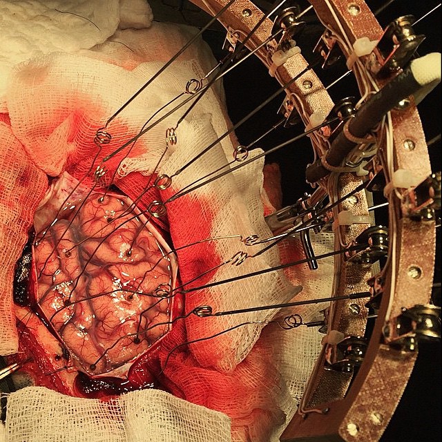
This is a picture of an intraoperative electrocorticography in a 28 year old male with refractory epilepsy that didn't have a clear epileptogenic lesion in the imaging studies. Refractory simply means an uncontrolled epilepsy (seizure) episodes, or ones that are resistant to anti-epileptic drugs. In such cases doctors need to map out the epileptogenic zone of the brain, to identify the location and limits of the area, and to decide if they can surgically remove it or not.
Electrocorticography is done by using electrodes placed directly on the exposed surface of the brain to record electrical activity from the cerebral cortex.

A mother hugs her newborn baby for the first time, the perfect photo to wish you all a happy Mother's Day! #happymothersday

Excision of a huge plexiform neurofibroma.
These are large and extensive benign tumors (non-cancerous) that grow from peripheral nerves anywhere in the body.
It involves single or multiple nerve fascicles that arises from major nerve branches and found in approximately 30% of patients with neurofibromatosis type 1 (NF1). Clinically, it presents as a cutaneous or subcutaneous mass (most commonly a superficial one), but can occur anywhere else.

Chin surgery!!! Genioplasty or the more common term known as chin repositioning, refer to the reduction and addition of material to a patient's chin. Basically using surgical implants that can alter the underlying structure of the face, providing better balance to the facial features.
Chin implants are used to build a better profile, using the patient's own bone taken from the ribs or from part of the pelvis.
This surgical procedure is done to correct receding chins, chin misalignment or chin excess.
The 28 years old male here had a convex facial profile and a 6 mm advancement genioplasty was the treatment plan for him.
The procedure presented in the photo was done and sent by Dr. Saeed Hajizadeh (@saeedhajizadehh), a maxillofacial surgeon, with the patient's full consent.

Surgical excision of a massive uterine fibroid!
Uterine fibroids (also known as leiomyomas) are a benign proliferation of smooth muscle cells of the uterus (myometrium), with the majority occurring during the childbearing years.
The interesting fact is that a single cell divides repeatedly, eventually creating a firm, rubbery tissue. They can grow slowly of rapidly, be single or multiple, and can vary in size from microscopic to the size of a full-term pregnancy!! They are classified by their location as follows - submucosal (beneath the endometrium), intramural (in the muscular wall of the uterus), and subserosal (beneath the uterine serosa - the outermost layer). Most women with fibroids have no clinical symptoms at all, but those who do, experience abnormal uterine bleeding which presents with menorrhagia (menstrual periods with heavy or prolonged bleeding), and pelvic pain.
Blood loss from fibroids can be heavy enough to cause chronic iron-deficiency anemia.
Depending on the size and the location, the fibroid can compress the adjacent organs and cause additional complications such as constipation, frequent urination and venous stasis.
There are theories that are believed to play a role in the cause of fibroids, these are hormonal changes (estrogen and progesterone) and genetics.
Very rarely, a cancerous fibroid can occur, known as a leiomyosarcoma.
Now the treatment is indicated only when there's a severe pain, heavy and irregular bleeding, infertility or pressure symptoms.
Basically there are medical therapies (GnRH agonists are decreasing the estrogen levels, and shrinks the fibroid), and a surgical treatment, myomectomy or hysterectomy.

Transplanted heart restarted after a shock with small paddles to restart the heartbeat and beating AV paced at 100 BPM. You can see the aortic arch stitch and the white wires are the pacing leads!
Source : Figure1

With the help of these guys the patient can confidently say to death "NOT TODAY!"

An incredible close-up shot of a living human brain. Seriously that brain is literally just showing off here!

When we're considering hernias the main concern can be strangulation.
Hernia can occur as a free sac without any content, but sometimes a loop of the intestine becomes stuck in the hernia (incarceration). Strangulation happens when the hernia traps the intestine so tightly that it cuts off the blood supply. The trapped part of intestine can develop gangrene in as few as just a couple of hours, causing extreme pain, tenderness of the area and symptoms of bowel obstruction. During gangrene, the intestinal wall dies, causing a rupture which leads to peritonitis (inflammation of the abdominal cavity) and further complications if left untreated. This condition is life-threatening and requires a surgical emergency.
In this photo sent by @r_grigoriu the ileum is pushed back to the abdominal cavity after clearing the strangulation.

Open reduction and internal fixation (ORIF) - part 2.
An intra-operative photo, shows the internal fixation of the broken bone using a metal plate and screws. It keeps the bone fracture stable in order to heal the right way and to help prevent infection.

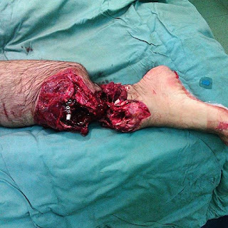
Virtually nothing is impossible in the world if you just put your mind to it and maintain a positive attitude, and this amazing girl (@bella_mj_) made it in every kind of sense. Her name is Izabela, she messaged me a couple of days ago, sending the bottom picture, and honestly she had a big impact on me after revealing the story hiding behind that photo.
Her story begins when she was 13 months old, she was burned in a fire that occurred in her bedroom while she was sleeping. She suffered 3rd and 4th degree burns all over her body, which caused sepsis and her both forearms were immediately amputated.
The bottom picture is of her second re-amputation of her lower leg, which was done on November 20th 2014, due to intensive bone growth of her lower leg after she had an amputation (June 25th 2012) and the first re-amputation (September 2012), which was done for the same reason. She asked the doctors to amputate it because she got sick of all the problems she had with it. Now, her fibula was removed and tibia shortened for 1cm.
Today she can use her abutments very good, which is the reason she refused to wear prosthetic hands. She can walk, run, dance, and everything a normal girl can do thanks to her prosthetic leg. She's starting to study medical laboratory diagnostics in a few months. Due to what she went through she started to love medicine and she's going to enroll medicine with the dream of choosing plastic surgery as her speciality. #respect

Open reduction and internal fixation (ORIF) following a fracture - Part 1.
ORIF refers to a surgical procedure to fix a severe bone fracture and is a two-part surgery. “Open reduction” means surgery is needed to realign the bone fracture into the normal position. “Internal fixation” refers to the steel rods, screws, or plates used to keep the bone fracture stable in order to heal the right way and to help prevent infection.
This surgery is done to repair fractures that would not heal correctly with casting or splinting alone.

This is a male patient, 19 years of age who had the two mandibular canines impacted in the chin by a deviation in their eruption routes, I.e. an ectopic movement of an unerupted mandibular canine.
Their removal was necessary to enable the other mandibular teeth to be orthodontic moved and the bite of the patient aligned.
The procedure was sent and done by @vladimirpoli_bucofacial

Do you know how a chronic subdural hematoma surgery is going?! Let's get into it, step by step!
Generally speaking, a chronic subdural hematoma is an "old" collection of blood and blood breakdown products between the surface of the brain and its outermost covering (the dura). It begins several weeks after the first bleeding.
So, from top to bottom, steps 1 and 2 shows the opening of the dura mater to reveal the hematoma's capsule; 3 - resection of the external layer of the hematoma; 4 - drainage of the blood clots inside the capsule of the hematoma, revealing the internal layer beneath it; 5 and 6 - resection of the internal layer and exposition of the brain and arachnoid.
This type of hemorrhage usually occurs in elderly people in use of anticoagulants or antiplatelet agents who suffer a head trauma, resulting in the damage of the bridging veins between the brain and dura mater, and slow accumulation of venous blood for weeks or months, eventually forming these capsules and clots compressing the brain to cause further damage.
Sent by a neurosurgery resident @rpaglioli

Yorumlar
Yorum Gönder