Yeni-New

Complete surgical removal of a dermoid cyst found in the floor of the mouth!
Dermoid cyst is generally benign and present at birth but are often diagnosed only later in life, and an association with pregnancy is commonly reported.
It's a sac-like growth that contains structures such as hair, fluid, teeth, or skin glands that can be found anywhere in the body, most commonly on the face, inside the skull, on the lower back and in the ovaries. They grow slowly and are not tender unless ruptured.
The best treatment is a surgical removal, superficial dermoid cysts (ones near the surface of the skin) on the face can usually be removed without complications. Removal of other, rarer dermoid cysts requires special techniques and training.
Credit to the maxillofacial surgeon @saeedhajizadehh

The human mind is so odd and creative that it hurts!
That's a bizarre X-Ray of a 10-centimeter dining fork, lodged in a 70-year-old man's penis!
This happened as a result of a strange and unlucky sexual accident. He arrived to the hospital and presented with bleeding from the area. The fork was not visible but doctors were able to feel it from the outside and X-rays showed its position.
You ask why? He told he had inserted the 10cm dining fork into his urethra almost 12 hours earlier in an attempt to achieve sexual gratification.
The doctors removed the item without any surgical intervention, in order to minimise urothelial trauma and preserve erectile function.

I never quite realized the true depth of this before, but after extensively studying the human body and its complex inner machinations, I can't help but feel so completely overwhelmed by how perfectly constructed we are.
This is a case of a spontaneous abortion (miscarriage) at around 19 weeks of gestation, which wasn't induced but happened spontaneously due to complications of the pregnancy.
As you look at it carefully, you realize and witness the true beauty of the human body even at gestational ages, how every part of our body syncs so flawlessly with one another and integrates with such mind-numbing complexity.

An enlarged heart, or cardiomegaly, is the term used to describe a heart that has a thickened wall or dilated chambers. Having a heart that is enlarged is not a disease but a symptom of an underlying medical condition.
This is a cardiomegaly at autopsy in a 27 year old who died from complications of morbid obesity. He weighted 423 pounds at his death. The normal male heart should fit in your hand, while this heart is 2-3x the normal size.
There's a direct association of adiposity with left ventricular dysfunction, independent of hypertension, coronary artery disease and other heart disease.
Obesity produces an increase in total blood volume and cardiac output because of the high metabolic activity of excessive fat. Eventually, the heart grows larger to compensate for the larger body.

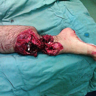
Foot fork-stabbing injury, better watch your steps!

Gunshot wound to the hand from a handgun fired at point blank range!
These days, gunshot injuries have become very common. Depending on the severity of the wound, appropriate measurements should be taken in the emergency department. The anatomy of the hand involves so many structure that an injury to an area may result in varying degrees of damage to different parts. The common hand injuries due to a gunshot are fracture on bones of the hand and fingers (the carpal bones, metacarpals, and phalanges), bone loss and instability, soft tissue injuries to the muscles, tendon and ligament injuries and damage to the nerve and blood vessels.
Following a limited debridement (removing dead tissue from the wound), the fractures are managed with early fracture stabilization, internal/external fixation, reduction and splinting. Follow-up surgeries are indicated depending on the extent and severity of the injury. Bone grafting is used in some cases, while in others amputation of digits or the damaged parts may be required. An infection during treatment should be managed with antibiotics.
The primary aim is to maximize functional ability and minimize the limitations or disabilities related to hand, which can be achieved with appropriate reconstruction and rehabilitation.

This is without a doubt the most touching one!
Sometimes doctors don't get involved with their patients, either by lack of time, or by the enormous number of patients we see each day. People use to think that surgeons or doctors in general are cold and have a bad bedside manner.
This photo proves the complete opposite, that apart from doing what we do for patients clinically, we can also offer a different treatment besides surgery or medications!
That little girl was operated for a brain tumor, she needed chemotherapy and couldn't be with other children in the hospital, so this fellow on the right (@rpaglioli - a current neurosurgery resident) brought his guitar and started signing songs with her, and her mother captured the moment.

Open Reduction and External Fixation (OREF) of an open tibial shaft fracture!
The tibia is considered as a subcutaneous bone, which makes it extremely vulnerable to open fractures, either by motor vehicle accidents, skiing accidents, or high-energy falls, as well as plenty of other causes.
OREF is usually necessary when a compound (open) fracture or if multiple bone fragments are present. It stabilizes bone and soft tissues at a distance from the operative or injury focus. The management of complex fractures is done with the help of an external fixator, this is a device used for injuries that are associated either with soft tissue damage or open wounds in the fracture area.
With an open reduction, the fractured ends of the bone are placed in alignment (the fracture is “reduced”), which promotes bone healing. With external fixation, pins are inserted through the skin into the bone and held in place by an external frame to keep it reduced (properly aligned), immobilized, or both.
In the above image, pins are inserted directly into the bone in this external fixation device used to realign broken bony parts.

The smallest little embryo with its amniotic sac!
This is a case of a miscarriage (or spontaneous abortion), which is the spontaneous loss of a fetus before the 20th week of pregnancy, or in this case even before the 12th week.
Keep in mind that miscarriage is a naturally occurring event due to some complications and has nothing to do with medical or surgical abortions, or any interruption of the pregnancy!
The common reasons pregnancies miscarry are chromosome problems, abnormal physical development or an abnormal placental development. Other causes can be infections, uterine problems, diseases of the mother (diabetes, lupus, thyroid disease), trauma, and unhealthy lifestyle (obesity, drug and alcohol use). The photo shows the embryo still with its intact amniotic sac and umbilical cord. The tiny black dot is the developing eye, and the reddish tissue in the center is the heart.

Pterygium growing onto the cornea!
People with pterygium have a triangular-shaped fleshy tissue that grows onto the cornea usually from the nasal side of the eye. It is a noncancerous lesion, usually not a serious condition that thought to be due to environmental factors such as ultraviolet light, chronic dryness and exposure to wind and dust.
Patients usually report on and off redness, mild discomfort, feeling of dryness or foreign body sensation and occasional tearing. Blurring of vision may be present if the pterygium encroaches too much onto the cornea causing astigmatism (irregular shape of the cornea) or if it covers the pupil.
Surgical excision, with or without conjunctival grafting (for reducing recurrence) is a common indication if the pterygium progresses towards the center of the cornea or for cosmetic reasons. During the surgery the pterygium is removed and the patient's own conjunctiva is either glued or stitched to the area in order to fill the empty space created by the removal of the pterygium.

Each of your feet has 26 bones, 33 joints, and more than 100 tendons, muscles, and ligaments. No wonder a lot of things can go wrong! This patient was lucky enough to have his foot back into one piece after a complex reconstructive surgery.
Reconstructive surgery of the foot and ankle consists of complex surgical repairs that may be necessary to regain function or stability, reduce pain, and prevent further deformity or disease. Unfortunately, there are many conditions or diseases that range from trauma to congenital defects that necessitate surgery of the foot and/or ankle.

There's a pretty classic GI condition for you to diagnose it!
Update - This isn't just a colon, it's a super duper megacolon!!! A megacolon denotes dilation of the colon that is not caused by mechanical obstruction. The common and useful measurement for diagnosing it is when the cecum is greater than 12cm (normally varies between 6-9 cm), the rectosigmoid region greater than 6.5 cm and the ascending colon greater than 8 cm.
A congenial megacolon is called Hirschsprung's disease, in which nerve cells of the myenteric plexus are absent, it typically affects babies, and therefore isn't the case here.
On the other hand, a toxic megacolon is a life-threatening complication that occurs in association with a severe attack of inflammatory bowel disease such as ulcerative colitis, Crohn's disease or an infection with Clostridium difficile. It is characterized by swelling and dilation of the large intestine due to inflammation and accumulation of excessive amounts of gas. Further complications like septic shock, hemorrhage and peritonitis rarely occur.
The goal of the treatment is to decompress the bowel and to prevent swallowed air from further distending the bowel. A colectomy (surgical resection of all of part of the colon) is usually indicated if the megacolon worsens.

Remember the fingertip injury? Here's another one! This was sent to me by @reyannalove shortly after posting the previous case.
She was tying up her horse and while she was doing that her horse reared on her which made the rope tighten and the rip go, caught around her finger and just popped it off. She underwent an emergency reconstructive surgery to her fingertip. Fortunately, she was lucky enough and her finger is now as good as new! In general, replantation or reconstruction of the fingertip when successful provides excellent cosmetic outcome by maintaining the digital length, preserving the nail and improving function.

Yorumlar
Yorum Gönder