 |
| Resim yazısı ekle |
If you think this cannot fit inside our bodies you're pretty much wrong!
Ovarian cysts are one of the things that surprise me the most. They are fluid-filled sacs or pockets within or on the surface of an ovary and are very common but rarely grow to mega sizes, generally harmless and disappear without treatment within a few months. They can cause symptoms like nausea, vomiting, bloating, painful bowel movements and pelvic pain.
To make things more complex, ovarian cysts are divided into functional and pathological ones. The functional develop as part of the menstrual cycle and are usually harmless and short-lived, while pathological occur due to abnormal cell growth, and are much less common.
This huge cysts seen above is a 10lbs taken from a middle-age female, notice how big it is compared to the surgeons hands!

Neurosurgery dose of the day!
Here's a glioblastoma multiforme (grade IV astrocytoma) from the right temporal lobe in a 56 years old female patient. This kind of tumor is the most common and most aggressive malignant brain tumor, it arises from astrocytes—the star-shaped cells that make up the “glue-like,” or supportive tissue of the brain and are highly malignant (cancerous) because the cells reproduce quickly and they are supported by a large network of blood vessels. The top images from the left shows T1 sagittal, T2 axial, and T2 coronal weighted MRI. The patient presented with progressive headache, left hemiparesis (remember right hemisphere has a control over the left body), and hyposthesia (reduce sense of touch). You may also notice the difference in the color and vessels from the healthy brain.
Treatment typically involves chemotherapy, radiation and surgery.

Here's a case you don't get to see every day, extensive facial trauma due to gas explosion!
Gas explosion is one of the most fearful accidents that happens due to a gas leak. People suffer from severe injuries that give even skilled surgeons some troubles.
Notice that the patients tongue is severed, the lips and a part of the mandible along with some teeth are detached from place and are being held by the surgeon. Clamps were placed on different areas to stops the bleeding, and a tracheal tube was inserted in order to assist breathing. This patient is alive and underwent a series of reconstructive operations in order to bring both aesthetics and functionality as close as possible to normal.

Approximately 7-8 week old aborted fetus due to a ruptured ectopic pregnancy.
As mentioned before, a pregnancy is ectopic when it occurs outside the womb (uterus). Ectopic means 'misplaced'. A ruptured ectopic pregnancy is one in which the fallopian tube gets torn or bursts and results in internal bleeding. Often there is intense abdominal pain, and sometimes, vaginal bleeding. In this case, laparoscopic surgery or abdominal surgery is usually needed, and either a part of the whole Fallopian tube along with the pregnancy will be surgically removed.

This photo shows a total laryngectomy on a 45 year old heavy smoker gentleman presenting with hoarseness of the voice and dysphagia. He was diagnosed with laryngeal squamous cell carcinoma and the treatment was surgical.
The larynx is your "voice box", the organ containing the vocal cords. The foremost risk factor for the development of laryngeal cancer is tobacco use. The risk of developing laryngeal cancer with tobacco increases with use and decreases after cessation.
Malignant tumors of the larynx may affect laryngeal physiology depending on tumor location and size. They can spread by direct extension to adjacent structures, by metastasis to regional cervical lymph nodes, or more distantly, through the blood stream.
Symptoms typically include hoarseness of the voice, persistent cough, a lump in the neck, stridor, and difficulty swallowing (dysphagia). Treatment depends on the location, type, and stage of the tumour and involve surgery, radiotherapy, or chemotherapy, alone or in combination. Laryngectomy (surgical removal of the larynx with the vocal cords) is done in severely affected patients.

This is a very interesting case of an ectopic pregnancy sent by arthur_trindade . The patient came to the emergency department with abdominal pain, characterized acute abdomen. The ultrasound revealed ectopic pregnancy and the gestational sac was removed.
An ectopic pregnancy occurs when a fertilised egg implants itself outside the womb. It most commonly occurs in a fallopian tube (this is known as a tubal pregnancy), usually as the result of damage to the fallopian tube or the tube not working properly. Signs and symptoms classically include abdominal pain and vaginal bleeding.
The pain may be described as sharp, dull, or crampy. Pain may also spread to the shoulder if bleeding into the abdomen has occurred.
If bleeding has already occurred, surgical intervention may be necessary. Surgeons use laparoscopy or laparotomy to gain access to the pelvis and can either incise the affected Fallopian and remove only the pregnancy (salpingostomy) or remove the affected tube with the pregnancy (salpingectomy).

Left hand laceration injury to a 27-year-old male who got into a fight!
Laceration is a cut, a deep cut in skin that is produced by the tearing of soft body tissue.
This type of wound is caused by a sharp object (such as a knife in this case or a shard of glass) and almost always leads to the injury of deeper structures under the skin such as vessels and nerves, and more rarely to tendons and bones.
The mechanism of injury can raise any suspicious of a nerve injury. In some cases, a simple bruise or swelling around the nerve will cause numbness or tingling for a few days. After this, normal feeling and function return. If the feeling doesn’t return within 10 days, the nerve has likely been cut.
A deep cut may be closed with stitches (sutures) or skin glue. Skin glue is used on cuts that have smooth edges and are not very deep and infected. This case requires surgical repair and application of stitches.
This is a case of a 5-year-old girl with Sturg-Weber syndrome that was operated last week.
Sturge-Weber syndrome is a rare disorder that is present at birth (congenital), characterized mainly by a congenital facial birthmark and neurological abnormalities. Other symptoms can include eye and internal organ irregularities. Each case of Sturge-Weber Syndrome is unique and exhibits the characterizing findings to varying degrees, so two patients coming with this condition will pretty much present different features.
The facial birthmark or "Port Wine Stain" present at birth and typically involving at least one upper eyelid and the forehead.
Neurological concerns relate to the development of excessive blood vessel growth on the surface of the brain (angiomas). These are located typically on the back (occipital) region of the brain on the same side as the port wine birthmark. These angiomas create abnormal conditions for brain function in the region.
This photo here clearly shows leptomeningeal angiomas on the young girl's right occipital and temporal lobes.

Scoliosis is a topic that interests me a lot, basically due to its treatment, surgical treatment. This is a side-view during scoliosis surgery.
Surgery is used to treat severe scoliosis. The goal of surgery is to improve a severe spinal curve. The result will not be a perfectly straight spine, but the goal is to balance the spine and to make sure the curve does not get worse. The most common type of surgery for scoliosis involves attaching rods to the spine and doing a spinal fusion. Spinal fusion is used to stabilize and reduce the size of the curve and stop the curve from getting worse by permanently joining the vertebrae into a solid mass of bone.
Other techniques are sometimes used, including instrumentation without fusion, which attaches devices such as metal rods to the spine to stabilize a spinal curve without fusing the spine together. This is only done in very young children when a fusion, which stops the growth of the fused part of the spine, is not desirable.
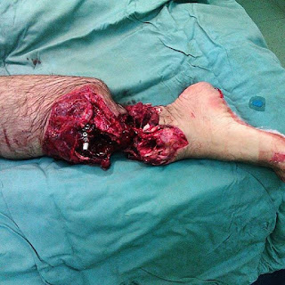
You see sometimes it's good to be fat, this male over here landed on wheat harvesting blade, after tripping on his shovel. Luckily it didn't get too deep and only severed his adipose tissue.
Median sternotomy for stab injury to left chest and ventricular laceration!
This patient was stabbed in the left anterior chest wall, 5th intercostal space, 1cm medial to midclavicular line. He was in shock with neck vein distension. FAST (Focused assessment with sonography for trauma) showed pericardial fluid, meaning that he was suffering from a ventricular wall laceration/tearing, and blood was leaking though it.
Ventricular repair, or cardiorrhaphy, has long been one of the most dramatic and lifesaving procedures performed in the emergency department.
In this photo, Index finger was placed on the actively bleeding site of injury and the laceration was repaired with 4-0 prolene via a pledget.


Yorumlar
Yorum Gönder